The A-band is the dark region of a sarcomere that spans the full length of the thick myosin filaments. It also includes the areas where thin actin filaments overlap with myosin at its edges, and it remains approximately the same length during muscle contraction.
What Is an A-Band in Muscle?
The A-band, short for anisotropic band, is the dark-staining region seen in striated muscle under a microscope. It appears dark because it contains thick myosin filaments, which refract light differently from the thinner actin filaments found in the lighter I-band.
The term “anisotropic” refers to the way the myosin filaments interact with polarized light, making this region appear darker than the surrounding areas. In simple terms, the A-band is the part of the sarcomere where you find the thick filaments.
In muscle physiology, an A-band refers to a specific region within a sarcomere, the primary muscle structure unit. The sarcomere comprises two central protein filaments, actin, and myosin, arranged in a highly ordered pattern to generate force and contraction.
The A-band is the dark region within a sarcomere that contains thick myosin filaments responsible for generating force during muscle contraction. The A-band also includes portions of thin actin filaments that overlap with the myosin filaments.
The length of the A-band remains constant during muscle contraction, while the size of the thin actin filaments changes. As a result, the overlap between the thin actin filaments and thick myosin filaments changes, allowing force and movement generation.
The A-band is named for its appearance in a microscope, where it appears as a dark band due to the high density of myosin filaments. The A-band is surrounded by lighter regions known as I-bands, which contain only thin actin filaments and no myosin filaments.
The size of the A-band can vary depending on the muscle type and the contraction stage. In general, longer A-bands correspond to stronger muscles, while shorter A-bands correspond to weaker muscles.
Overall, the A-band is a critical component of muscle physiology, and its structure and function play a crucial role in generating force and movement.
Understanding the properties and behavior of the A-band is essential for understanding muscle function and developing treatments for muscle-related disorders.
Location and Structure of A-Band in Muscles
To understand where the A-band sits, first we have to look at how muscle is organized from large to small. Skeletal muscle is made up of muscle fibres, and each fibre contains repeating units called sarcomeres. The A-band is located inside the sarcomere, right in its central region.
A muscle fibre is a single muscle cell. Inside each muscle fibre are long, thread-like structures called myofibrils. These myofibrils are made up of repeating units called sarcomeres, arranged end to end like beads on a string.
Each sarcomere is the basic functional unit of muscle contraction and is defined as the segment between two Z-lines, which act as its boundaries.
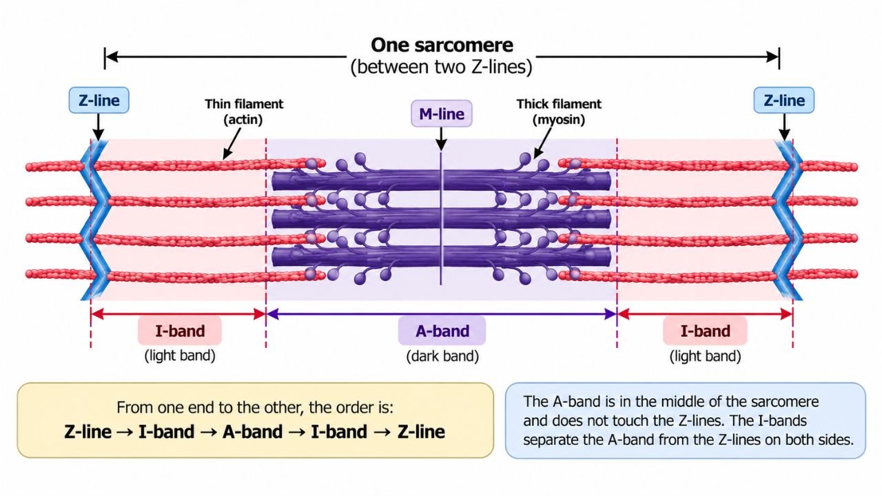
If you look at one sarcomere from one end to the other, the order of structures is Z-line, then I-band, then A-band, then I-band, and then Z-line again. This means the A-band sits right in the middle of the sarcomere, with one light I-band on each side of it.
The A-band does not touch the Z-lines because the I-bands are in between, separating the A-band from the Z-lines on both sides.
The A-band is located exactly where the thick myosin filaments are present. It begins where the thick filaments begin and ends where they end, which is why it occupies the central part of the sarcomere.
The thick myosin filaments are anchored at the M-line in the middle of the sarcomere and extend outward from there, so the A-band naturally sits in the central region, flanked by I-bands on both sides.
Inside the A-band itself, there are two important sub-regions. The H-zone lies at the centre of the A-band and contains only thick myosin filaments, with no actin overlap. This is why the H-zone appears slightly lighter than the rest of the A-band, where actin and myosin overlap at the edges.
The M-line runs through the middle of the H-zone, right at the centre of the sarcomere. It is made of proteins that hold the thick myosin filaments together and anchor them in place. The “M” in M-line stands for “middle,” because it is located at the exact midpoint of the sarcomere.
The most important thing to remember is that the A-band corresponds exactly to the length of the thick myosin filaments. Wherever myosin is present, that is part of the A-band. This is why the A-band is in the centre of the sarcomere.
The myosin filaments are anchored at the M-line in the middle and extend outward, so the A-band naturally sits in the central region, flanked by I-bands on both sides.
In short, the A-band is the dark band located in the centre of each sarcomere, between the two I-bands. It contains the full length of the thick myosin filaments, with actin filaments overlapping at its edges, and the H-zone and M-line are found inside it at the centre.
Function of the A-Band in Muscles
The A-band plays a central role in muscle contraction because it contains the myosin filaments that interact with actin to generate force. However, it is important to understand that the A-band alone does not produce contraction.
The process involves the entire sarcomere and the regulated interaction between actin and myosin filaments.
Myosin has small projections called cross-bridges (or myosin heads) that attach to specific sites on the actin filaments. When these cross-bridges attach and pull, they generate the force that causes muscle contraction.
Role of the A-Band During Muscle Contraction
Muscle contraction is explained by the sliding filament theory. This theory says that muscles shorten because thin actin filaments slide past thick myosin filaments, pulling the Z-lines closer together.
Step-by-step process:
- Nerve signal arrives: A motor neuron sends a signal to the muscle fibre.
- Calcium is released: Calcium ions are released inside the muscle fibre.
- Cross-bridges attach: Myosin heads on the thick filaments attach to binding sites on the thin actin filaments.
- Power stroke: The myosin heads pull the actin filaments toward the center of the sarcomere.
- Filaments slide: Actin filaments slide past myosin filaments, pulling the Z-lines closer together.
- Sarcomere shortens: The sarcomere becomes shorter, which means the muscle contracts.
What changes and what stays the same During the Muscle Contraction?
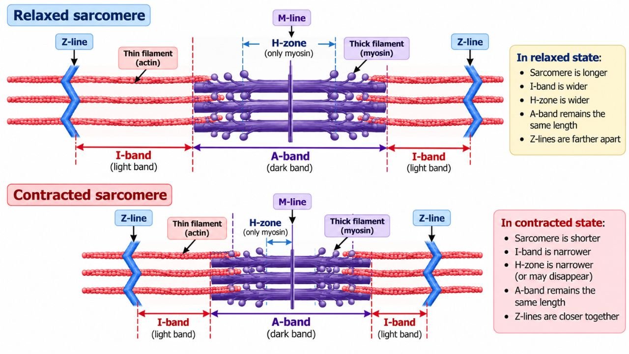
During contraction:
- The A-band stays the same length because the myosin filaments themselves do not shorten.
- The I-band becomes narrower because actin filaments slide further into the A-band.
- The H-zone becomes narrower because actin filaments move into the region that previously had only myosin.
- The Z-lines move closer together as the sarcomere shortens.
A-Band vs. I-Band vs. H-Zone
This comparison is one of the most useful revision tools for students.
| Feature | A-band | I-band | H-zone |
| Location | Central region of sarcomere | On either side of the A-band | Middle of the A-band |
| Filaments present | Thick myosin + overlapping thin actin at edges | Only thin actin filaments | Only thick myosin filaments |
| Appearance | Dark | Light | Lighter than the rest of the A-band |
| Length during contraction | Stays the same | Becomes narrower | Becomes narrower |
| Contains M-line? | Yes, at the center | No | Yes, at the center |
Why Does the A-Band Remain Unchanged During Contraction?
The A-band remains the same length during contraction because the thick myosin filaments themselves do not shorten. Instead, the thin actin filaments slide past them, pulling the Z-lines closer together.
This is the key idea of the sliding filament theory: filaments slide past each other, but the filaments themselves do not shorten. Because the myosin filaments keep their original length, the A-band, which spans their full length, also keeps its original length.
In contrast, the I-band and H-zone become narrower because actin filaments slide deeper into the A-band, reducing the regions where only one type of filament is present.
Frequently Asked Questions
Q: Where is the A-band located?
The A-band is in the central region of the sarcomere and corresponds to the full length of the thick myosin filaments.
Q: What is the A-band made of?
It is made of thick myosin filaments, with thin actin filaments overlapping at its edges. The H-zone in the middle contains only myosin.
Q: Why does the A-band appear dark?
It appears dark because the myosin filaments are anisotropic, meaning they interact with polarized light in a way that makes this region darker than the I-band.
Q: What happens to the A-band during muscle contraction?
The A-band stays the same length because the myosin filaments do not shorten. Only the I-band and H-zone become narrower as actin slides past myosin.
Q: What is the function of the A-band?
It contains the myosin filaments whose cross-bridges interact with actin to generate the force needed for muscle contraction.
Q: What is the difference between the A-band and the H-zone?
The A-band is the entire dark region containing myosin (with actin overlap at the edges). The H-zone is only the middle part of the A-band where there is no actin overlap, just myosin.
Key Points for Exams
- The A-band is the dark region of the sarcomere containing thick myosin filaments.
- It spans the full length of the myosin filaments and includes actin overlap at its edges.
- The H-zone is inside the A-band and contains only myosin.
- The M-line is at the center of the A-band and anchors the myosin filaments.
- During contraction, the A-band stays the same length, while the I-band and H-zone become narrower.
- This happens because actin filaments slide past myosin filaments, not because the filaments themselves shorten.
Short self-test
1. Which filament type is found in the A-band?
Answer: Thick myosin filaments.
2. Does the A-band get shorter during contraction?
Answer: No, it stays the same length.
3. What is the H-zone?
Answer: The middle part of the A-band where only myosin is present, with no actin overlap.
4. What anchors the myosin filaments in the A-band?
Answer: The M-line.
5. Why do the I-band and H-zone narrow during contraction?
Answer: Because actin filaments slide deeper into the A-band, reducing the regions with only one filament type.
References
- NCERT Biology, Chapter 17: Locomotion and Movement: https://ncert.nic.in/textbook/pdf/kebo117.pdf
- OpenStax Anatomy and Physiology, “Muscle Fiber Contraction and Relaxation”: https://openstax.org/books/anatomy-and-physiology/pages/10-3-muscle-fiber-contraction-and-relaxation
- OpenStax Biology 2e, “Muscle Contraction and Locomotion”: https://openstax.org/books/biology-2e/pages/38-4-muscle-contraction-and-locomotion
- NCBI Bookshelf, “Actin, Myosin, and Cell Movement”: https://www.ncbi.nlm.nih.gov/books/NBK9961/
- PMC, “Muscle Contraction”: https://pmc.ncbi.nlm.nih.gov/articles/PMC5793755/